

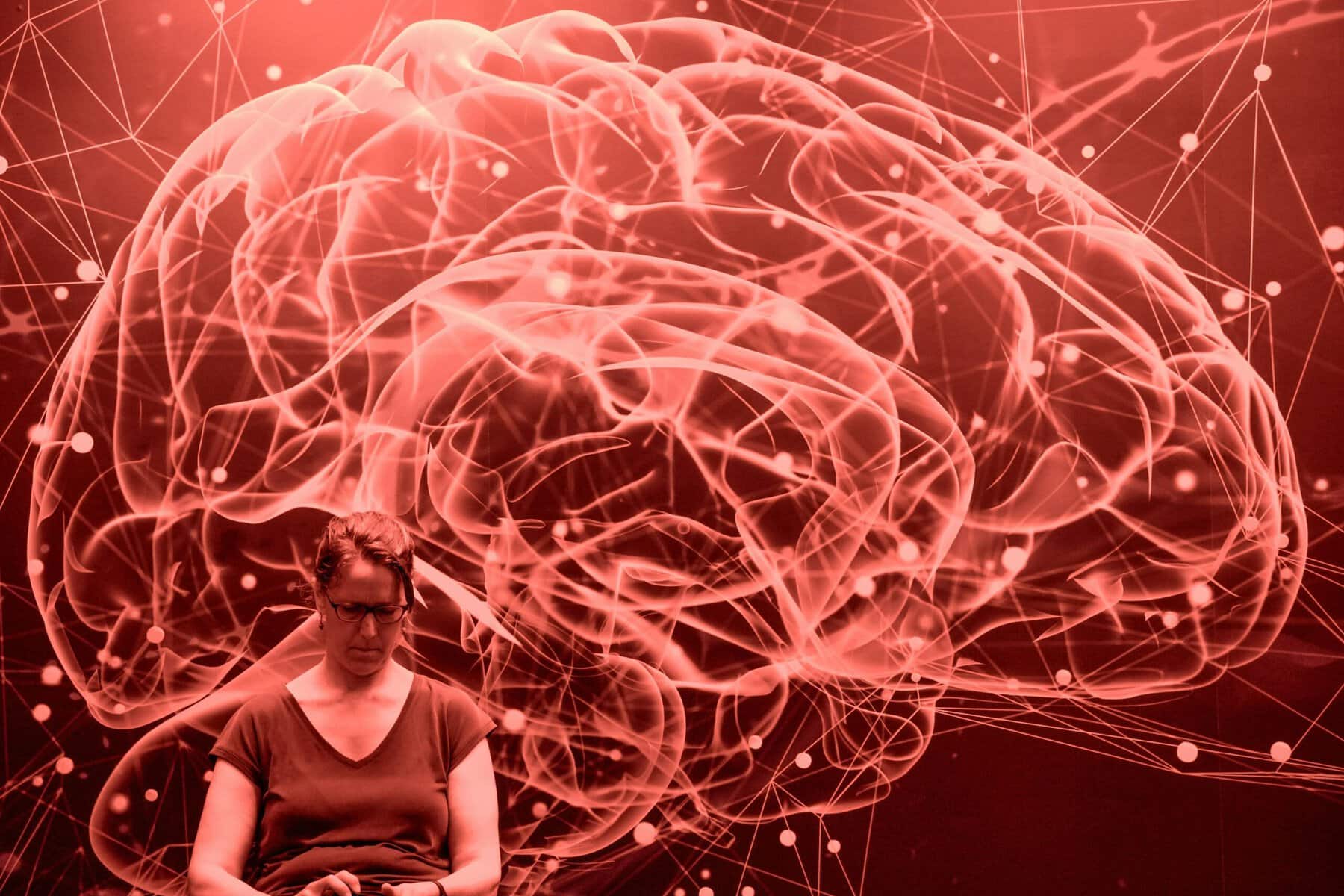


The human brain is extraordinarily complex, what with 80 billion neurons or more, each often connected with thousands of other nerve cells.
Even so, much has been learned, especially in the last two centuries, about what parts of the brain do what, especially for the simpler components of the brain such as the sensory and motor systems.
Their stratified anatomy and physiology lend themselves to study much easier than cognitive functions, feelings and emotions or, for some scientists, solving the riddle of the nature of consciousness.
The last challenge bedevilled some of the best scientists in the 20th century, such as the physicist Francis Crick (Nobel Prize winner, 1962), who tried for several decades to make sense of consciousness. Even now, the study of consciousness is dogged by widely differing, competing hypotheses.
There was hope in those early days: Crick suggested that light might be the solution to deciphering the highly complex and fast electrical signaling characteristic of the mammalian brain.
This may be so, given recent developments in optogenetics studies, which were rewarded by this year’s Nobel Prize in medicine or physiology to Karl Deisseroth, Peter Hegemann and Georg Nagel for their pioneering work in the field of optogenetics.
Early work on photosensitive single algae cells revealed that a light stimulus of the correct frequency could trigger an electrical signal in photosensitive receptors in the outer membranes of these cells.
Soon the genes responsible for those receptors were identified and later inserted into cultured frog nerve cells. This allowed investigators to stimulate nerve cells with light
The next step was obvious: insert light-sensitive genes into nerve cells in the region of a rat’s brain that control movement of the whiskers, then stimulate those cells with light signals using a fine light probe inserted into the selected region of the rat’s brain. That too worked.
The approach was elegant because specific types of nerve cells in the brain could be stimulated or inhibited depending on the type of receptor and light employed. Since then, the method has been used to study memory engrams and emotions such as fear.
But elegant as the method is, it was never designed to understand the development of the brain. For that purpose, another method was needed.
John Gurdon (Nobel Prize winner, 2012) in the U.K. showed that nuclei from mature fully differentiated columnar cells in the tadpole gut could be transplanted into a frog embryonic cell whose nucleus had been destroyed and the embryonic cell would go on to develop into a tadpole — even an adult frog.
Shinya Yamanaka (Nobel Prize winner, 2012) working in Japan showed much the same: cultured adult mouse fibroblasts treated with four transcription factors developed into pluripotent cells capable of being redirected to develop into adult mice.
Together, what these studies showed is that mature cells retain all the genes of the embryonic stem cell and have the ability to differentiate into any cell in the body given the right redirecting molecular signals.
For example, embryonic stem cells could be redirected to create parts of the brain such as the neocortex and highlight in detail the exquisitely timed multiplication, differentiation, migration and contact patterns of normal embryonic development including when specific genes were turned on or off during development of embryonic layered neocortex.
Here is the perfect model for studying the earliest stages of normal in utero development or disorders such as autism or schizophrenia that might affect the early development of the brain.
There are, however, limitations.
Those several-millimetre “minibrains,” as they are referred to, have no connections to other parts of the brain or other tissues, the absence of which could inhibit or disturb development of the brain and, with no blood supply, minibrains depend entirely on diffusion for food and oxygen and clearance of the waste products of development.
Still, it’s quite an advance and could be linked with optogenetics to study communication between developing cells by seeing who’s wired to what nerve cells to do what.
Going further, parts of the brain such as the pallidum, cerebellum, or even spinal cord can be developed in much the same way and, if brought together, form contacts with one another much as they might normally and form what some investigators call assembloids.
Development of the brain is busiest in the embryo and first few years of life when most connections are made and winnowing of connections not needed or otherwise faulty are cleared up without leaving a trace. By the third decade, development has mostly come to an end.
The combination of minibrains and optogenetic methods could shed light on many diseases, genetic or otherwise in early life by studying when the first molecular, physiological and pathological changes take place and whether intervention at the earliest stages might prevent some disorders from developing.
To study the development and maturation of the brain is to be reminded how vulnerable the developing child’s brain could be to exposure of the mother to drugs or alcohol when development is so finely tuned and easily derailed.
The same is true during childhood, adolescent and even early adult years when the brain continues to develop and might be easily set back by malnutrition, harmful drugs or physical trauma, to say nothing of psychological trauma.
Dr. William Brown is a professor of neurology at McMaster University and co-founder of the InfoHealth series at the Niagara-on-the-Lake Public Library.